
Introduction to Quantum Ophthalmology
Source: arXiv:2606.19238 · Published 2026-06-17 · By Mukhit Kulmaganbetov, Dmitry Pushin, Taranjit Singh, Pinki Chahal, David Cory, Iman Salehi et al.
TL;DR
This survey paper explores the emerging intersection of quantum technologies and ophthalmology, highlighting four key directions: photon-limited retinal imaging, correlation-based (ghost) imaging, nanoscale quantum dot probes, and quantum-limited studies of visual perception. The paper details advances in optical coherence tomography (OCT) enhanced by single-photon detection that enable imaging under strict photon budget constraints, thus reducing retinal phototoxicity while preserving image quality. Quantum optical coherence tomography (Q-OCT) using entangled photon pairs offers proof-of-principle improvements via nonclassical correlations, though practical clinical adoption is nascent. Correlation-based ghost imaging, including three-dimensional versions with single-photon avalanche diodes (SPADs), provides alternative low-light imaging paradigms but currently suffers from limited detection efficiency and acquisition speed. Nanoscale quantum dots (QDs) with quantum confinement effects offer highly photostable, size-tunable fluorescent probes suitable for enhanced contrast and targeted drug delivery, yet face unresolved clinical translation challenges such as toxicity and biocompatibility. Finally, the authors review psychophysical and theoretical studies probing human visual system responses at the single-photon level and to structured light fields carrying orbital angular momentum (OAM), elucidating how human vision operates near physical detection limits and can serve as a biological detector of quantum states. Overall, the paper argues quantum and quantum-inspired methods present promising new tools for ophthalmic imaging and vision science but remain in early developmental stages with multiple technical hurdles to clinical implementation.
Key findings
- Single-photon detector-based OCT systems achieve high-quality retinal images at incident photon fluxes as low as ~10 pW, significantly reducing phototoxic risk while maintaining image quality comparable to conventional OCT [55].
- Quantum OCT (Q-OCT) using entangled photon pairs can resolve multi-layer retinal samples via interferograms where dips correlate with sample optical thickness, demonstrating proof-of-principle quantum advantage in axial resolution [57].
- On-chip spectral domain OCT implementations enable in vivo retinal imaging at eye power levels around 480 µW to 830 µW, showing feasibility of photonic integration to miniaturize OCT systems for clinical use [58].
- Three-dimensional ghost imaging microscopes using time-correlated single-photon detection with orthogonal microscope objectives enable volumetric imaging without mechanical scanning at low light levels [78].
- All-digital ghost imaging combined with neural networks can reconstruct super-resolved images from entangled photon pairs under sparse illumination, pointing toward data-driven noise suppression strategies [77].
- Quantum dots exhibit size-tunable emission spectra and greater photostability than conventional dyes, facilitating longitudinal imaging with enhanced contrast, though concerns remain about toxicity and biocompatibility [96–99, 117].
- Human rod photoreceptors can detect individual photons with significant probability above chance, and psychophysical experiments show perception of classical structured light (e.g., spin-orbit coupled fields) modulated by orbital angular momentum states [29, 30–32].
- Advanced quantum-inspired stimuli reveal classical analogs to entanglement through human visual perception of spatially and polarization-nonseparable light fields, opening avenues for noninvasive optical probing of retinal structure [142].
Threat model
The threat model is primarily practical and biomedical rather than adversarial: imaging and probing the retina and visual system under extreme photon budget constraints to avoid phototoxicity, with limitations posed by photon flux, detector noise, and biological safety requirements. There is no active attacker; instead, the challenge is to maximize image quality and diagnostic capability at the quantum limit of light exposure.
Methodology — deep read
The paper is a broad review synthesizing multiple experimental approaches and theoretical concepts rather than reporting a single new study. The threat model is generally biomedical: imaging and probing visual function under limits of photon exposure to avoid phototoxicity, considering photons as scarce quantum resources. The adversary here is practical constraints such as low photon budgets, detector noise, and biological safety rather than active attackers.
Data samples cited include retinal images from prototype OCT setups operating under low photon flux (approx. 10 pW), experiments producing entangled photon pairs via spontaneous parametric down-conversion for Q-OCT, and optical stimuli with structured light (OAM) applied to human subjects for psychophysics. Details on dataset sizes are sparse due to the review nature; references point to individual experimental papers.
Architectural descriptions cover various optical configurations: a Linnik–Michelson interferometer paired with superconducting single-photon detectors (SSPDs) and FPGA-based timing electronics for photon-resolving OCT measurements; SPDC photon-pair interferometers with beam splitters for Q-OCT; integrated silicon photonic platforms incorporating broadband sources, waveguides, and arrayed waveguide gratings for on-chip spectral domain OCT; 3D ghost imaging microscopes with orthogonally oriented objectives and SPAD arrays for coincidence photon measurements; and quantum dot nanoparticles engineered with tunable emission spectra and surface functionalization for targeted imaging and therapy.
The training regime primarily pertains to computational methods such as neural networks applied to reconstruct and denoise ghost images from sparse photon detections, but no concrete ML training details (epochs, batch sizes) are provided here. Most experiments involve physical optical setups rather than machine learning models.
Evaluation protocols focus on comparing image quality, signal-to-noise ratios, and spatial/axial resolution under constraints of photon flux, detector efficiency, and acquisition speed. Psychophysical protocols include threshold detection assessments of single photons and stimuli varying OAM quantum numbers to quantify perceptual responses.
Reproducibility is limited by early-stage, bespoke experimental setups. Several cited works provide code or detailed methods, but many datasets are proprietary or require specialized quantum optics hardware. Quantum dot synthesis and functionalization protocols vary across studies with limited standardization.
One example: In the single-photon OCT setup [55], a pulsed laser is attenuated to low photon number and directed into a Linnik–Michelson interferometer where one arm interacts with the retinal sample. The recombined photons pass through a dispersive fiber spool acting as a spectrometer before detection by an SSPD with precise time-stamping. This allows reconstructing spectral interferograms to form depth-resolved images at ultralow input powers (~10 pW), much lower than classical OCT. FPGA electronics timestamp photon arrivals enabling high temporal resolution; the system achieves image quality comparable to conventional OCT without risking phototoxicity due to low illumination. The approach demonstrates feasibility of quantum-inspired detection improvements.
Overall, the review carefully discusses the interplay of novel quantum optical sources, detectors, computational methods, and biological systems to explore new frontiers in ophthalmic imaging and visual science while emphasizing practical hardware and biological constraints.
Technical innovations
- Use of superconducting single-photon detectors combined with dispersive Fourier transform and FPGA time-stamping to achieve photon-limited OCT with drastically reduced illumination power [55].
- Implementation of quantum OCT leveraging entangled photon pairs for correlation-based interferometric imaging that achieves axial thickness measurement beyond classical limits [57].
- Development of integrated photonic chip-based spectral domain OCT systems with on-chip arrayed waveguide gratings enabling compact, stable retinal imaging at clinically safe illumination levels [58].
- Application of time-correlated single-photon detection in orthogonal optical paths to realize three-dimensional ghost imaging microscopes allowing volumetric reconstruction without scanning [78].
- Combining all-digital ghost imaging with neural networks to enable super-resolution image reconstruction under sparse entangled photon illumination patterns [77].
Datasets
- Low-photon OCT retinal data — ~10 pW photon flux images — Experimental data from [55]
- Quantum OCT interferograms — SPDC entangled photon pairs — Demonstrated in [57]
- On-chip OCT retinal images — In vivo human subjects at 480-830 µW power — From silicon photonic platform [58]
- 3D ghost imaging datasets — Correlated photon timing from SPAD arrays — Experimental proof-of-principle [78]
- Psychophysical visual perception datasets — Human detection of single photons and structured light perception — Various referenced works [29, 30–32, 142]
Baselines vs proposed
- Conventional OCT: image quality at standard illumination power vs single-photon detector-based OCT: comparable image quality at ~10 pW, dramatically reduced phototoxicity [55]
- Classical interferometric OCT vs quantum OCT (Q-OCT): Q-OCT yields interferograms resolving optical thickness dips with higher axial discrimination, though acquisition speed remains slower [57]
- Standard fluorescent dyes vs quantum dots: QDs show substantially higher photostability and tunable emission enabling longer-term imaging [96–99]
- Standard scanning OCT vs 3D ghost imaging microscope: ghost imaging achieves volumetric data without mechanical scanning under low illumination, though with trade-offs in acquisition time [78]
- Standard visual stimuli vs structured light with OAM: enhanced classical entoptic visual patterns observed with OAM stimuli indicating perception of spatial-polarization nonseparability [142]
Figures from the paper
Figures are reproduced from the source paper for academic discussion. Original copyright: the paper authors. See arXiv:2606.19238.
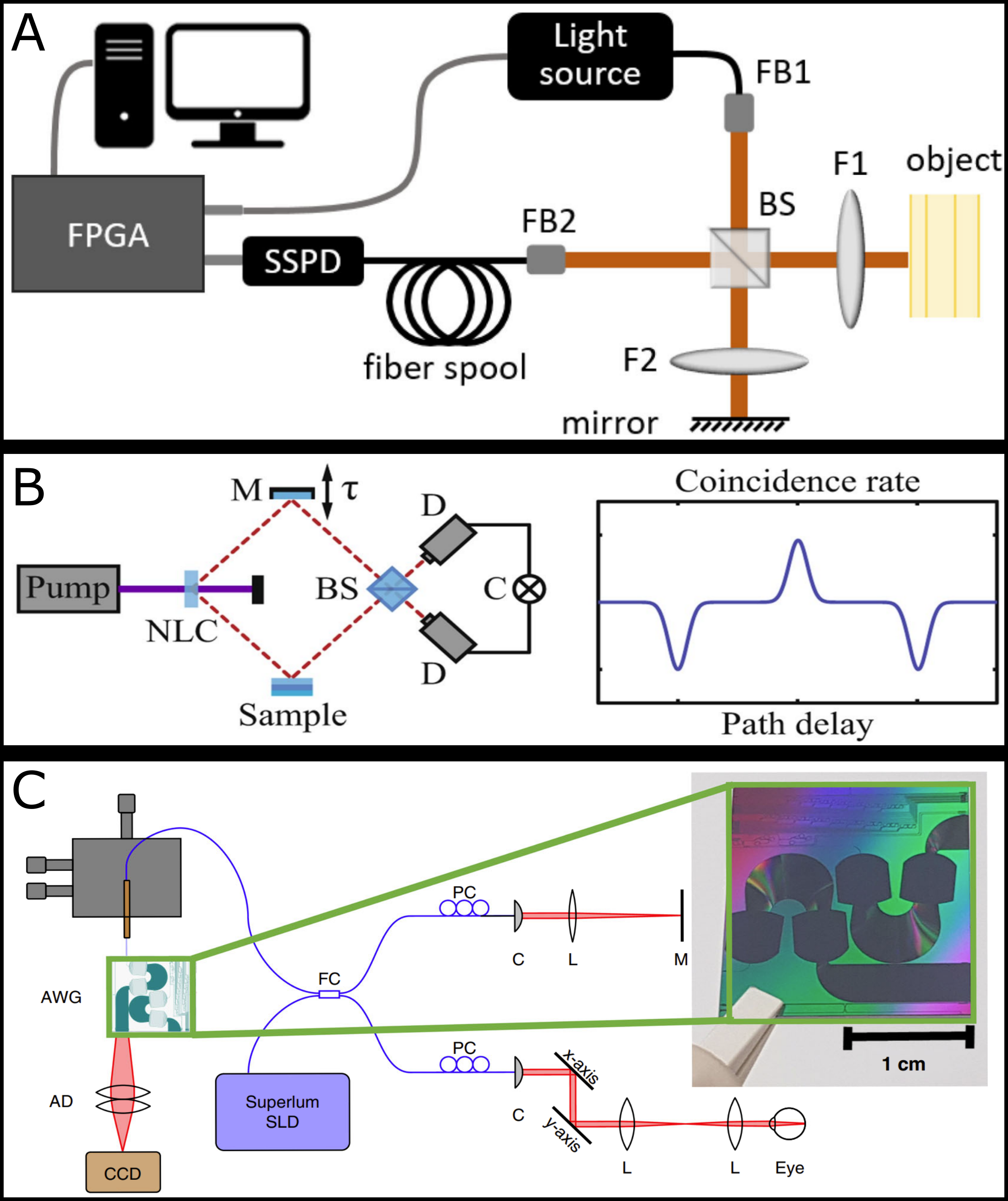
Fig 1: Advanced optical coherence tomography (OCT) modalities for ad-
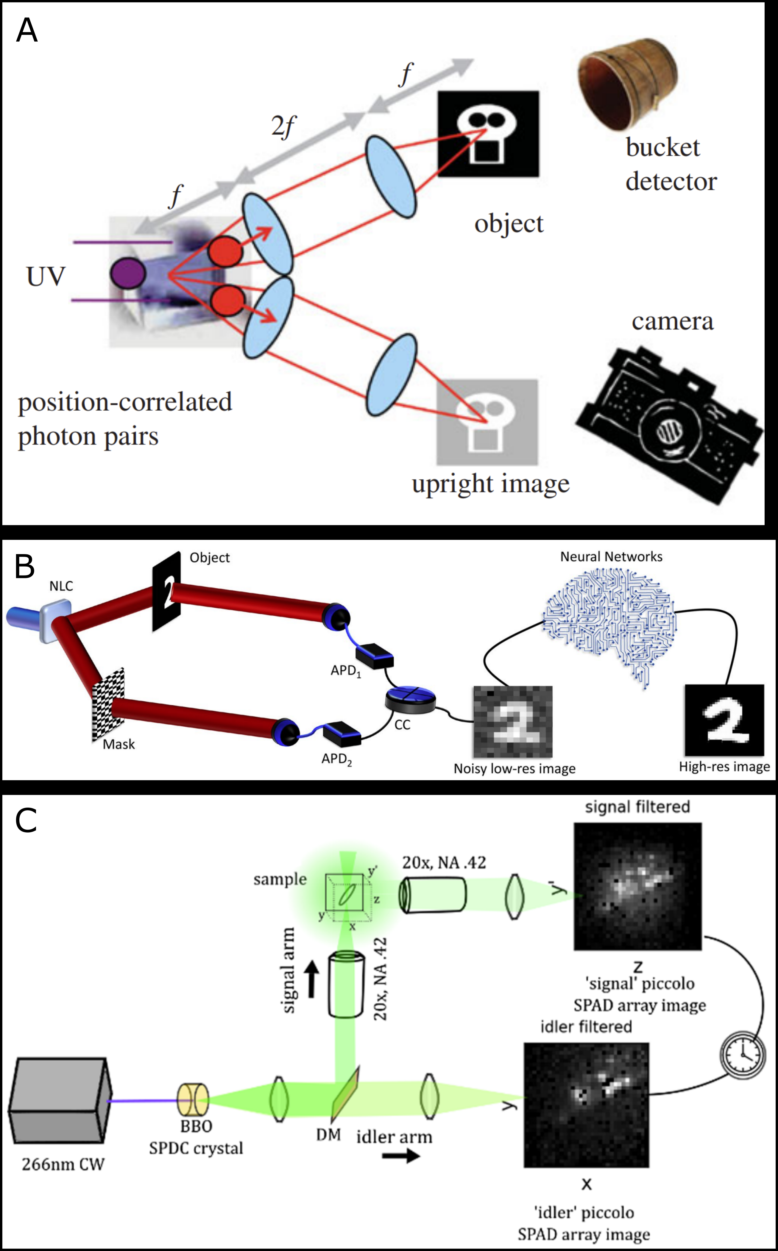
Fig 2: Advances in quantum and computational ghost imaging for volu-
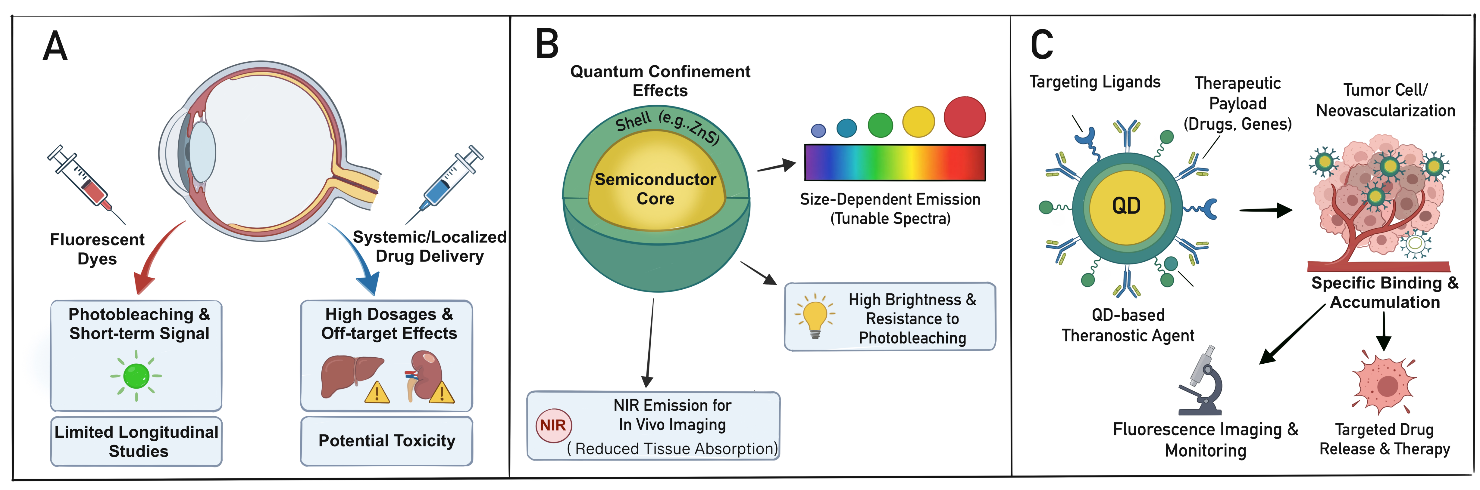
Fig 3: Schematic overview of quantum dots (QDs) for ophthalmic imaging
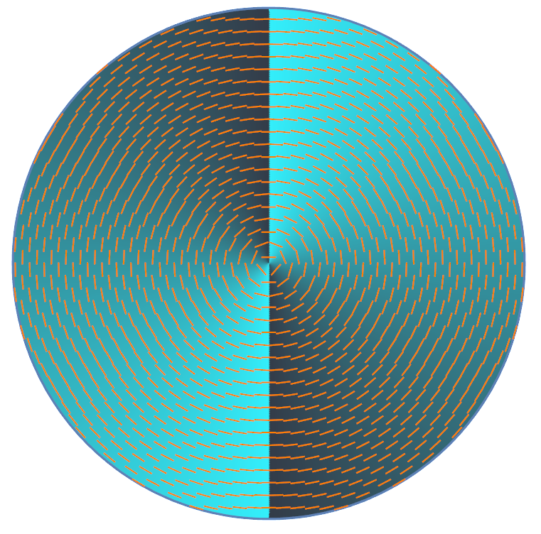
Fig 4: Examples of the phase and polarization patterns of structured light

Fig 5 (page 12).
Fig 6 (page 12).

Fig 7 (page 12).

Fig 8 (page 12).
Limitations
- Most quantum-enhanced ophthalmic imaging methods are at proof-of-principle stage and not yet integrated into routine clinical workflows.
- Quantum OCT systems face challenges including slow acquisition speeds, system complexity, and requirements for high detector efficiency.
- Ghost imaging approaches exhibit limited practical utility due to low detection efficiency and long measurement times incompatible with real-time imaging.
- Quantum dots pose significant unresolved concerns over long-term toxicity, biocompatibility, clearance pathways, and regulatory hurdles preventing near-term clinical adoption.
- Human vision quantum perception studies are limited by experimental complexity, small subject cohorts, and challenges separating quantum effects from biological noise.
- Integration of quantum light sources, detectors, and photonic circuitry into compact, robust devices suitable for clinical ophthalmology remains an open engineering challenge.
Open questions / follow-ons
- Can quantum OCT systems be optimized to achieve clinically relevant acquisition speeds and robustness?
- What are scalable methods to improve detection efficiency and reduce measurement time for ghost imaging in ophthalmic contexts?
- How can quantum dots be engineered to ensure safe long-term biocompatibility for human ophthalmic use?
- To what extent can structured quantum or classical light stimuli be leveraged for sensitive clinical diagnostics probing retinal microstructure and visual function?
Why it matters for bot defense
For bot-defense and CAPTCHA practitioners, the paper offers insight into ultra-sensitive quantum-limited photon detection technologies and optical correlation imaging approaches that push the limits of low-light sensing. Analogously, CAPTCHA systems may draw inspiration from quantum- or photon-level sensing to detect subtle automated interaction signals or spoofing attempts that rely on low signal levels or quantum-inspired randomness. The discussed quantum detectors and correlation-based imaging could inform development of CAPTCHAs incorporating physical unclonable functions or optical noise signatures difficult for automated solvers to reproduce. Furthermore, the analysis of human perception at single-photon and structured light levels underscores the difficulty of replicating biological-level detection heuristics, a principle that can be leveraged for designing human-interactive tests resistant to machine simulation. However, all these quantum-enabled methods remain experimental and complex, so their direct application to bot defense would require significant engineering simplification and cost reduction. Nonetheless, the fundamental limits and quantum-inspired sensing outlined provide conceptual avenues for novel CAPTCHA modalities exploiting photon-level measurement and spatiotemporal correlation properties inaccessible to bots.
Cite
@article{arxiv2606_19238,
  title={ Introduction to Quantum Ophthalmology },
  author={ Mukhit Kulmaganbetov and Dmitry Pushin and Taranjit Singh and Pinki Chahal and David Cory and Iman Salehi and Andrew Silva and Ben Thompson and Dusan Sarenac },
  journal={arXiv preprint arXiv:2606.19238},
  year={ 2026 },
  url={https://arxiv.org/abs/2606.19238}
}